

#ScienceSaturday posts share exciting scientific developments and educational resources with the KAND community. Each week, Dr. Dominique Lessard and Dr. Dylan Verden of KIF1A.ORG summarize newly published KIF1A-related research and highlight progress in rare disease research and therapeutic development.
KIF1A-Related Research
Lattice light-sheet microscopy and evaluation of dendritic transport in cultured hippocampal tissue reveal high variability in mobility of the KIF1A motor domain and entry into dendritic spines
Have you ever noticed that neurons look a little… funky? One can use the analogy comparing neuron morphology, or shape, to the shape of a tree by breaking it down into three parts. We often discuss the “trunk” (axon) of neurons where KIF1A-mediated axonal transport occurs towards the “roots” (synaptic terminals) to send messages to other cells in the nervous system. But today we are discussing a different part of the neuron: the dendrites, or “branches”. The dendrites are a neuronal structure where input or information is received from other cells. Like branches on a tree, dendrites also have “leaves” called dendritic spines. These dendritic spines help the neuron gather information from its surroundings.
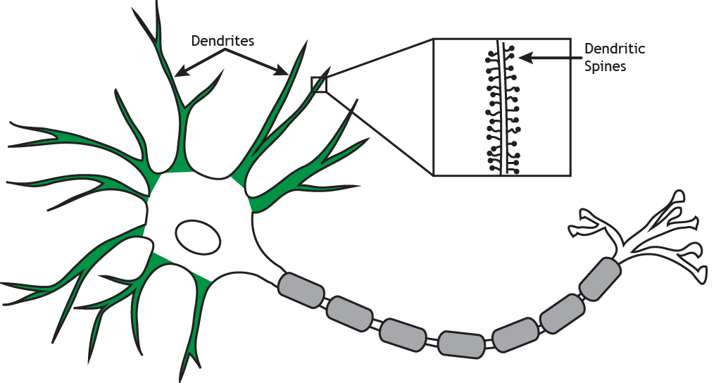
Molecular motor proteins, such as KIF1A, play an important role in the development and maintenance of dendritic branches by delivering and removing specific cellular cargo to precise locations within the dendrite. In the paper we are sharing today, authors did an in-depth investigation of KIF1A’s location and movement in and out of the dendrites of neurons. Specifically, KIF1A movement was visualized in “CA1 hippocampal pyramidal” neurons in a slice of mouse brain! This study identified that in these neurons’ dendrites, KIF1A movement is variable:
- KIF1A engages in long smooth continuous movements in one direction.
- KIF1A also engages in short segments of movement with many directional changes.
- Surprisingly, KIF1A spends a lot of time stalled, or stationary, inside of the dendrites.
The authors concluded that KIF1A tends to choose a dedicated direction to move, even though it could switch directions in dendrites.
How are scientists able to visualize intricate KIF1A movement inside of cells? Furthermore, how can this information be gathered inside of a piece of brain tissue? The answer lies in astounding technical advancements in microscope engineering! While the first microscopes emerged around the 17th century, today’s modern microscopes look vastly different.
Vision Engineering.
In the context of this paper, a technique called volumetric lattice light-sheet microscopy is used to image neurons in slices of mouse brain tissue. This allows for very fast imaging of neurons with impressive clarity, without damaging cells in the process. To watch light-sheet microscopy in action, have a look at the video below!
Rare Roundup
Rare Parents Tackling Rare Diseases
Parents are a relentless force in the rare disease space, driving the advancement of treatment, community, advocacy, and more. In this article you will learn about three parent pioneers in the rare disease space, focused on community building, facilitation, and drug development. We are continuously amazed by the power of rare disease parents – the many roles they play while navigating the world of rare disease is astounding and we thank them for their leadership.
“While Casey, Mike, and Effie have made their mark finding their niches in advancing the science and awareness of rare diseases, they are not alone. Countless other parents and family members have also taken up the mantle of pushing for cures, funding for research, securing payer reimbursements, and providing help to navigate the healthcare system. These tireless parents are the new face of advancement in treating rare disease and prove that there is strength in numbers. While one parent can make an impact, it’s the community of rare patients that will continue to accelerate progress in the field.”